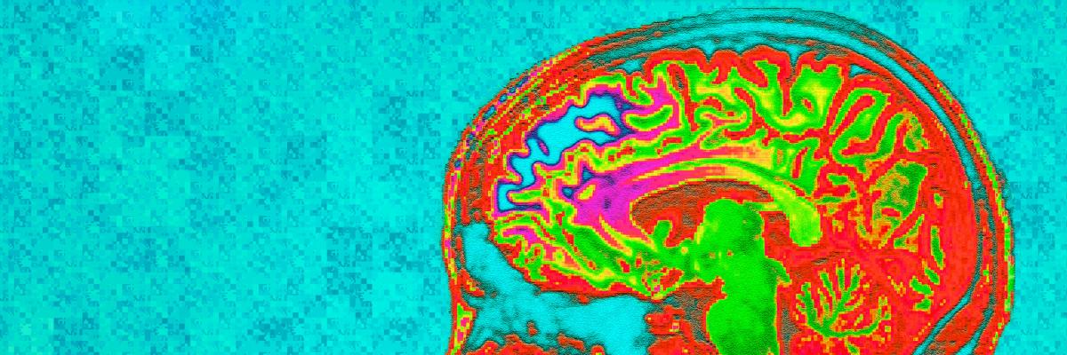
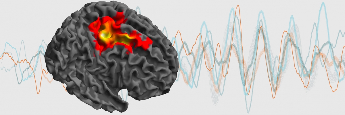
Rendered whole brain magnetic resonance image showing which cortical areas have their activities coupled with those in the motor region of the subthalamic nucleus in Parkinson’s disease. Coupling is determined by simultaneous recordings of magnetoencephalographic activity and deep brain recordings in patients with electrodes implanted for deep brain stimulation treatment. Representative deep brain waves are also shown. The effects of drugs and of high frequency stimulation on coupling and deep brain activity can thereby be determined, and provide clues as to how treatments can be improved.