Schools Open Day 2026
Schools Open Day 2026
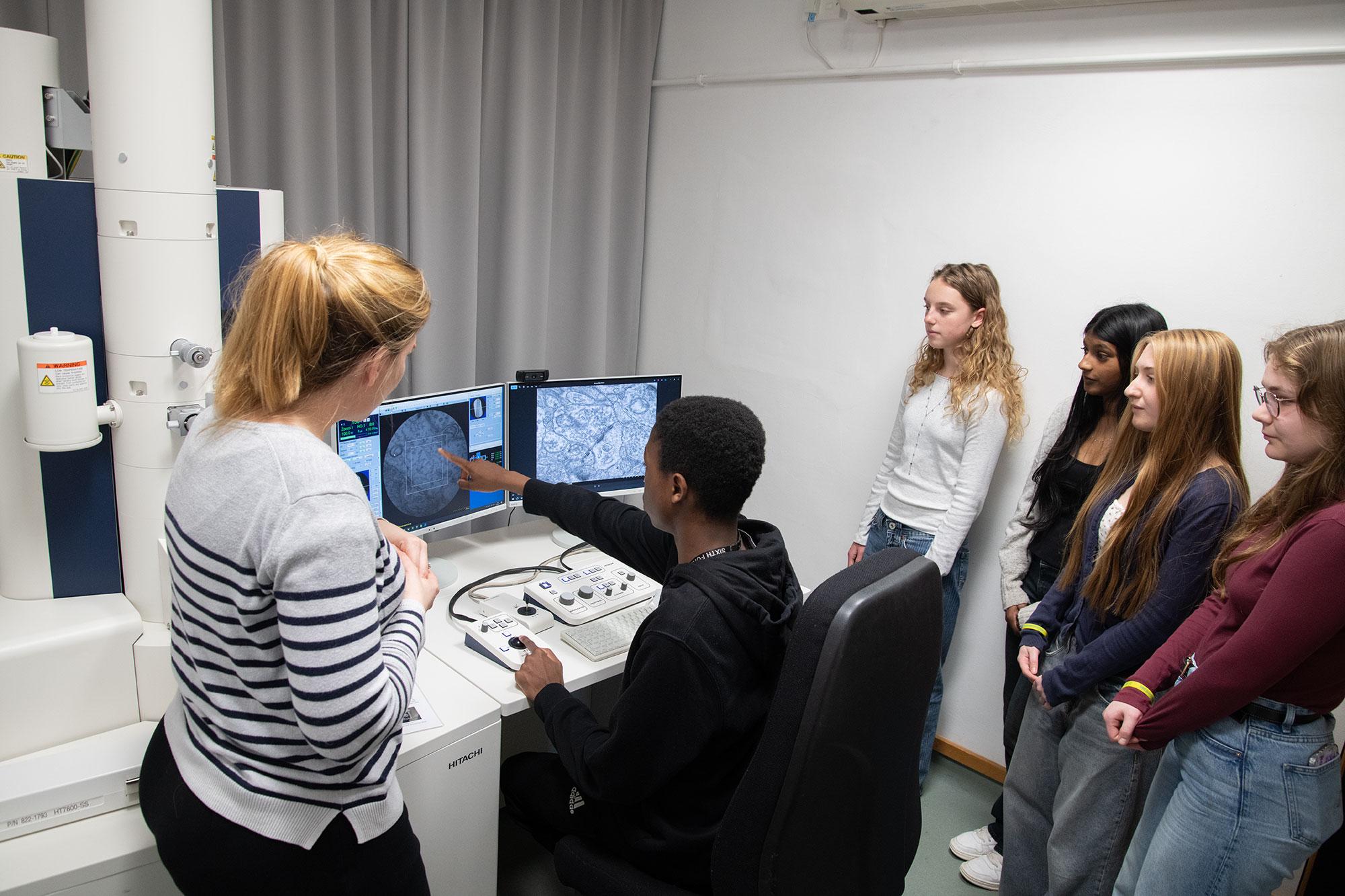
Dr. Natalie Doig (left) answers questions from pupils as they get a chance to explore the smallest structures of brain cells for themselves, using the Unit’s transmission electron microscope
The Brain Network Dynamics Unit was delighted to once again host groups from local state-funded schools for its annual Schools Open Day, held this year on 19th March.
Unit staff and students welcomed pupils and teachers to learn more about STEM (Science, Technology, Engineering and Mathematics) and medical research at the Unit. The Open Day started with a showing of “The Symphony of the Brain” video to familiarise visitors with the Unit’s research. Some of the Unit’s early-career researchers then talked about their experiences of working in STEM, highlighting their diverse educational backgrounds and career paths that led them to join the Unit. This was followed by an interactive discussion of the use of animals in research.
Pupils then rotated in small groups through different hands-on activities led by more early-career researchers. Special emphasis was placed on giving pupils the opportunity to see real working instruments and laboratories for themselves. Engagement activities were coordinated around four ‘knowledge stations’, at which pupils could learn about the Unit’s key research themes, including the activity and structure of the brain in health and disease; human brain stimulation; computer modelling of brain function; and brain-machine interfaces. The visit ended with a wrap-up session capturing feedback.
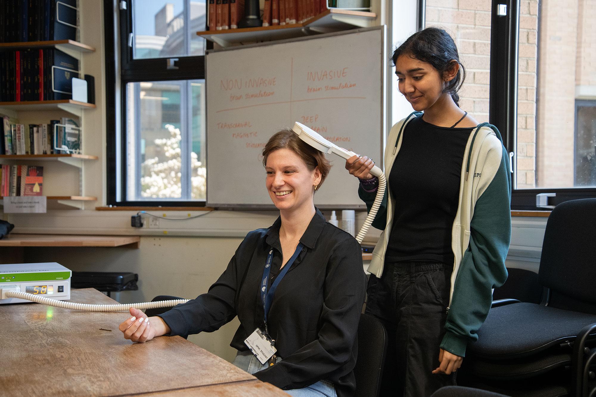
Dr. Camille Lasbareilles guides a pupil to the right part of her motor cortex for hand control. Using transcranial magnetic stimulation, Camille’s hand twitches when the stimulator is correctly placed.
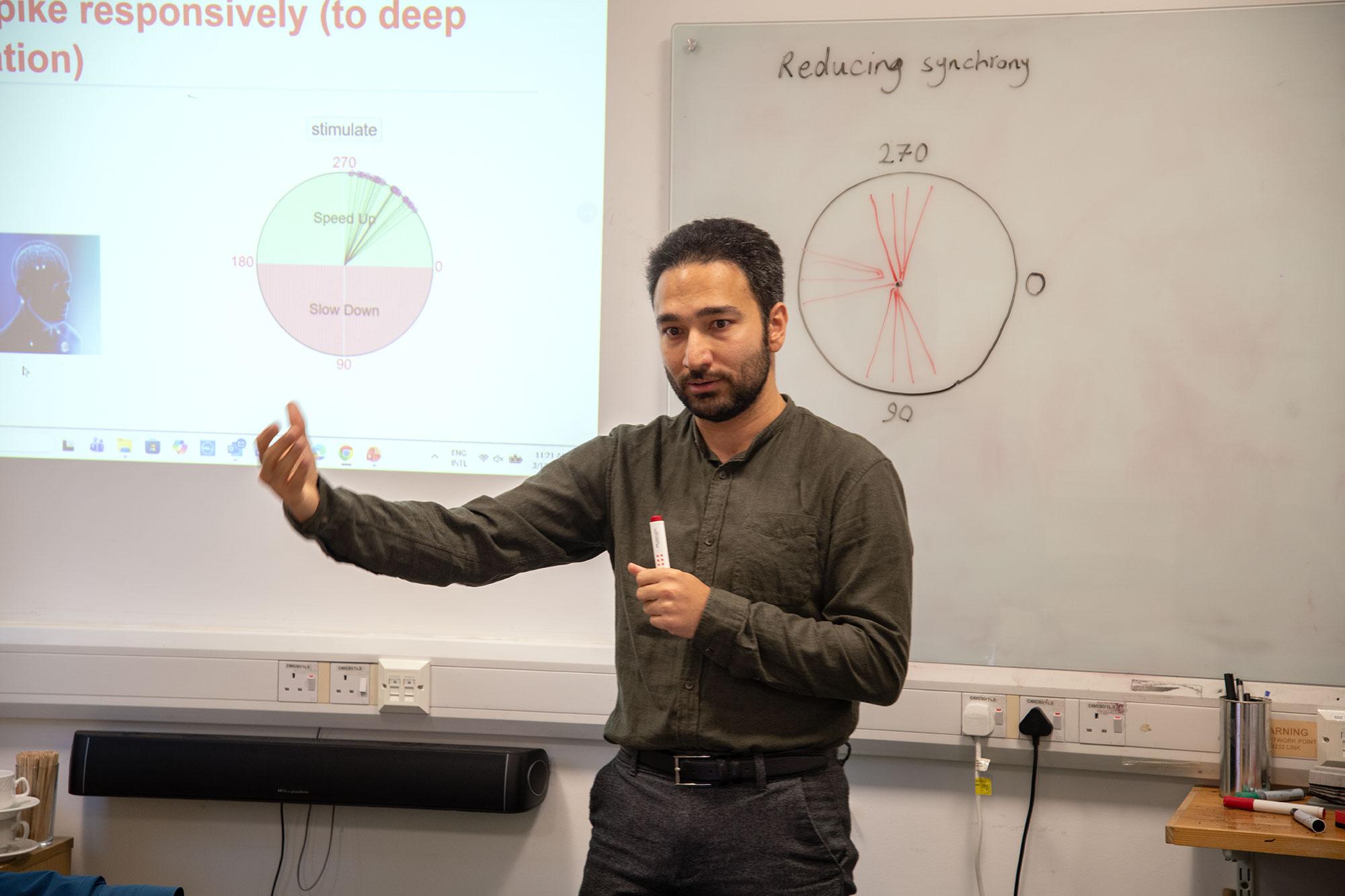
Dr. Nima Mirkhani discusses students’ strategies to secure a ‘high score’ in a game designed to explain deep brain stimulation optimisation to disrupt excessively synchronised neural activity.
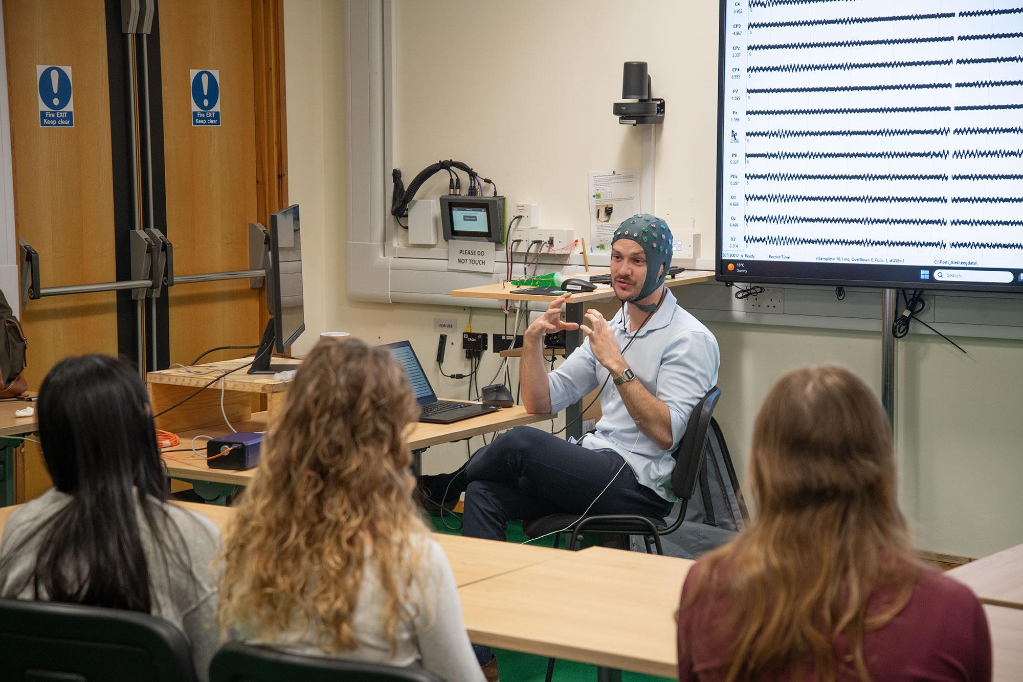
Dr. Fernando Rodrigeuz Plazas explains EEG brain recording- with a live readout from his own EEG is shown on the display behind him.
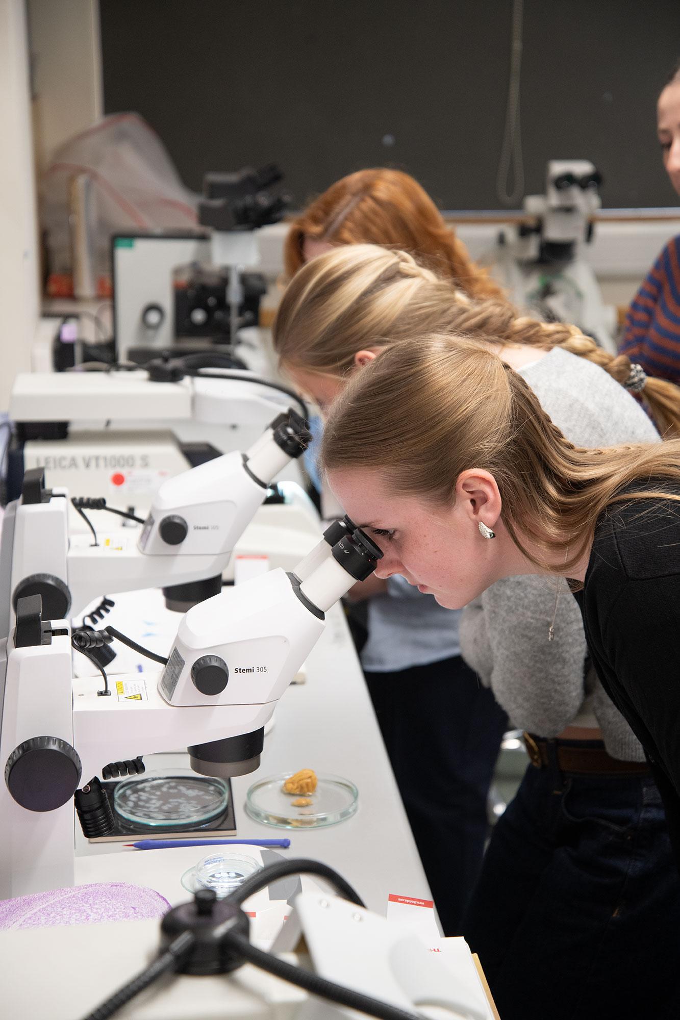
Visiting pupils see a working histology lab and follow the workflow for brain tissue from an intact brain, through sectioning and immunolabelling, to mounting on slides.
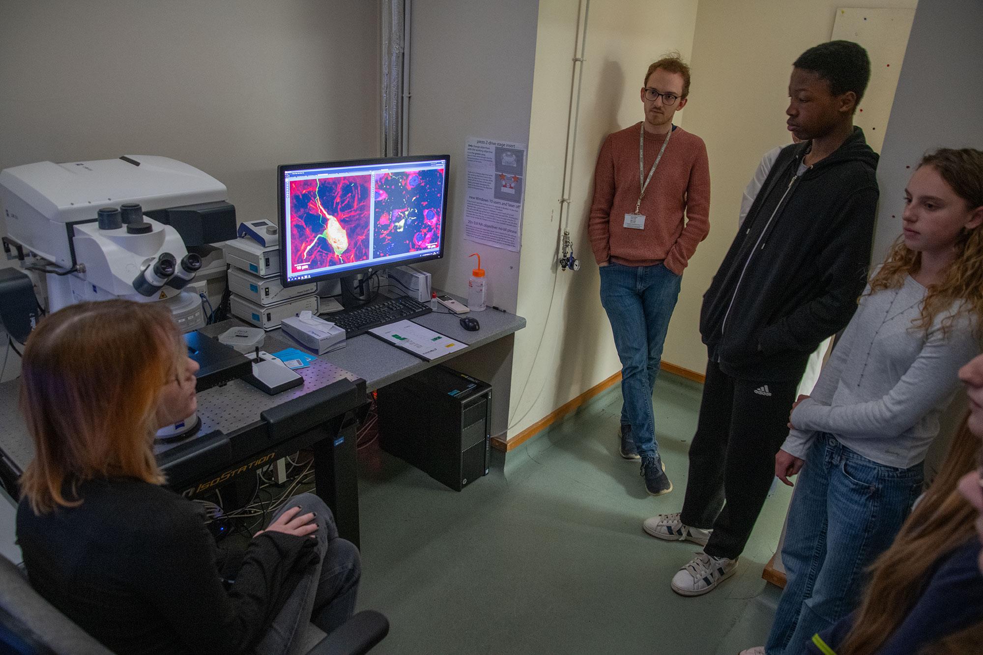
Jennifer Blackmore explains to pupils and their teacher how the Unit’s laser scanning confocal microscope allows them to see the coloured labels that have been applied to parts of brain cells- through transgenic markers, or antibody labelling.
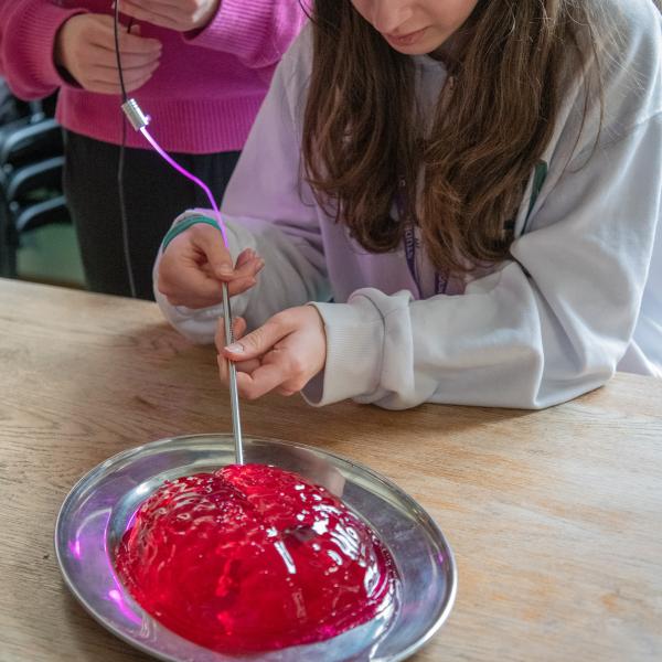
A visiting school pupil tries their hand at implanting a dummy stimulation electrode in a jelly brain!

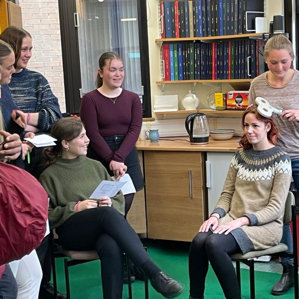
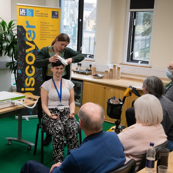
Unit early-career researchers Camille Lasbareilles and Ioana Grigoras demonstrate transcranial magnetic stimulation to members of the Unit’s Patient and Public Advisory Group.